Technology Platform

From baby tooth to patient-specific neuron

Our DPSC platform converts shed deciduous teeth into functional neuronal models of rare disease — without reprogramming, in a fraction of the time.

Why DPSC

A fundamentally better starting material

Induced pluripotent stem cells (iPSCs) require viral transduction of Yamanaka factors to reprogram somatic cells back to a pluripotent state — a process that takes 60–84 days, introduces epigenetic artifacts, and costs $1,500+ per vial before differentiation.

Dental pulp stem cells (DPSC) are neural crest-derived multipotent stem cells that reside inside deciduous teeth. Because they share a developmental lineage with neurons, they differentiate directly into functional cortical-like neurons without any transgenic reprogramming — in 7–14 days for initial DPSC expansion, and mature into neurons in 6–7 weeks. Critically, DPSC differentiation obviates any transgenic expression, retaining cellular naivety and giving researchers an open canvas to develop and test new ASO, CRISPR, and other therapeutic modalities without a reprogramming background.

Critically, DPSC retain disease-relevant epigenetic marks. They more closely resemble embryonic stem cells than iPSCs, making them superior models for imprinting disorders like Angelman syndrome, Prader-Willi, and Dup15q.
Key Advantages
No reprogramming No viral transduction required. Start neuronal differentiation immediately.
7–14 day DPSC expansion Neurons mature in 6–7 weeks vs. 60–84 days for iPSC. Faster iterations, faster science.
Biological replicates Multiple lines per genotype. Real statistical power.
Epigenetic fidelity Preserves imprinting marks critical for chr. 15 disorders.
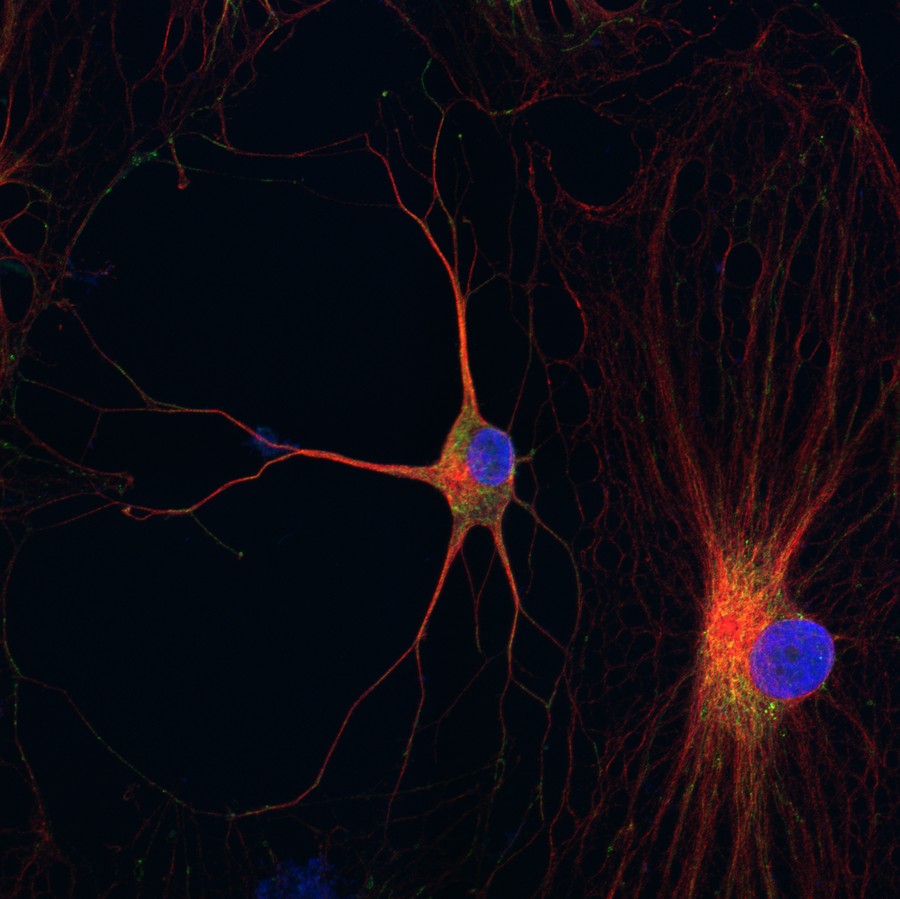
DPSC-derived neurons showing dendritic morphology
DPSC-derived neurons (Beta-tubulin / TOMM20 / DAPI). Mature cortical-like morphology at 4 weeks. Reiter Lab.
Differentiation Protocol

3-step neuronal differentiation

Our validated protocol reproducibly generates cortical-like neurons expressing MAP2, GABA-A, Neuroligin, and functional Na⁺/K⁺/Ca²⁺ ion channels.

1
Tooth Collection
Baby tooth, shipped by parents
2
DPSC Expansion
Isolate & culture stem cells
3
Epigenetic Reset
5-azacytidine treatment
4
Neural Induction
NTF + PKC/cAMP activators
5
Maturation
6–7 weeks in culture
6
Assay Ready
MEA, circadian, drug screening

Reference: Goorha & Reiter (2017). Curr Protoc Hum Genet. PMID: 28075485

Assay Capabilities

Functional readouts that matter to drug developers

MEA Electrophysiology

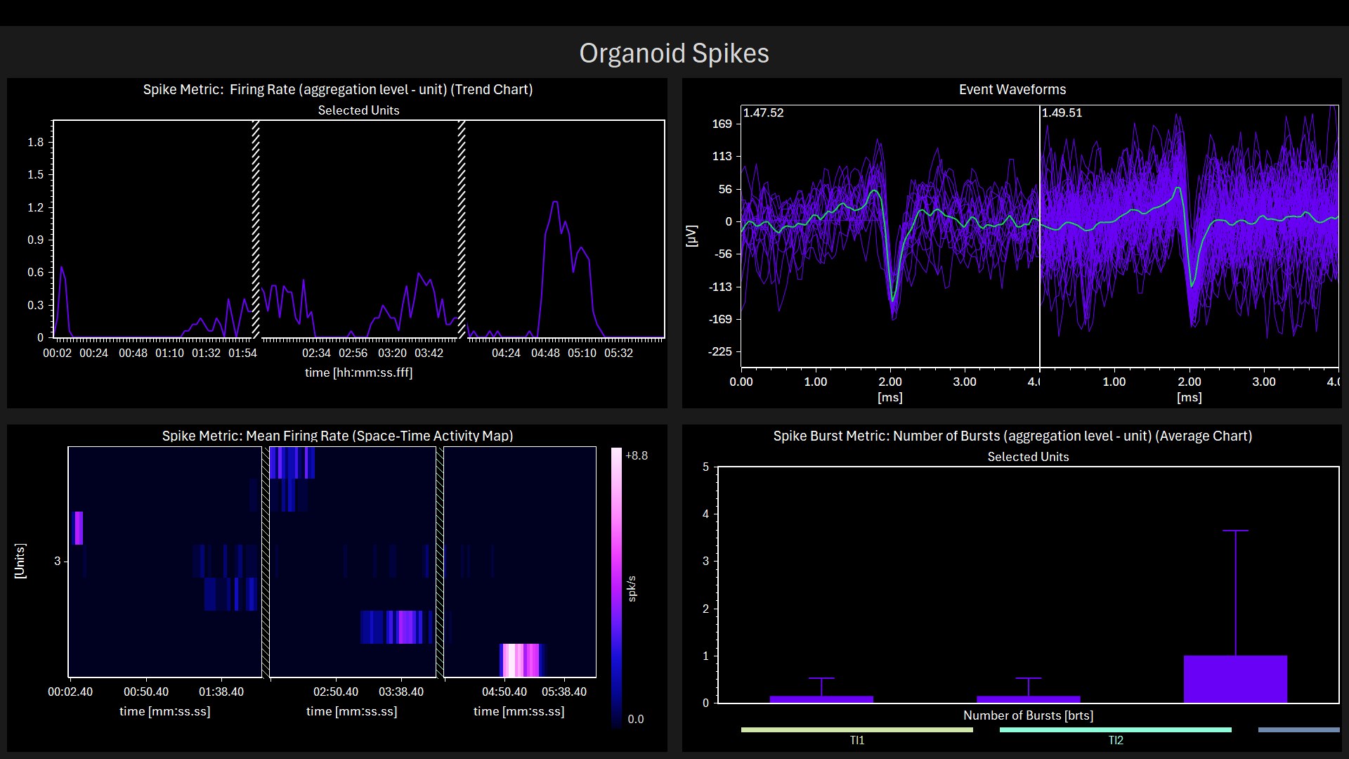
Multielectrode array recordings from DPSC-derived brain organoids capture firing rate, burst patterns, and network synchrony. 4-AP seizure induction validated.

Rodriguez TC et al., in preparation
🕐

Circadian Rhythm Assays

Per2:luciferase reporter system in DPSC neurons enables 5-day LumiCycle recordings of circadian period, amplitude, and phase. Validated in Prader-Willi neurons.

Victor AK et al. (2025). HGG Advances. PMID: 40023766
🧬

Transcriptomics & Methylome

RNA-seq, ATAC-seq, and WGBS profiling of disease vs. control neurons. Demonstrated for Dup15q, Angelman, and PWS — revealing novel disease mechanisms.

Urraca N et al. (2018). Mol Autism. PMID: 29423132
💊

ASO Therapeutic Screening

Antisense oligonucleotide validation across multiple patient cell lines per genotype. Test ASO efficacy and toxicity in the neuronal background where the therapy will act.

Applicable to Angelman syndrome, Dup15q, and other UBE3A/imprinting targets
🔬

Small Molecule Screening

Reporter constructs (luciferase, fluorescent) enable high-throughput compound screening. Circadian-correcting drugs, seizure suppressants, and mitochondrial rescue compounds.

Platform currently in development
✂️

CRISPR-Cas9 Gene Correction

DPSC neurons serve as the test bed for CRISPR-based UBE3A reactivation (Angelman) and other gene correction strategies in patient-specific neuronal backgrounds.

Angelman UBE3A target in development
3D Models

DPSC-derived brain organoids

Beyond 2D neuronal cultures, PulpNeuro is developing DPSC-derived brain organoids — 3D tissue models that better capture the network complexity relevant to epilepsy and seizure biology.

Neuronal organoids are measured by MEA, enabling real-time recording of spontaneous and evoked activity. 4-AP challenge confirms epileptiform activity in disease-relevant genotypes.

Organoid Development Team

Dr. Tyler Rodriguez — Neuronal organoids & MEA electrophysiology

MEA recordings from DPSC-derived neuronal organoids
MEA spike metrics from DPSC neuronal organoids. Firing rate (top left), event waveforms (top right), space-time activity map (bottom left), burst count (bottom right). 4-AP seizure induction shown.
Competitive Landscape

PulpNeuro vs. iPSC alternatives

Technology Disease Lines Cost/Vial Time to Neurons
✦ PulpNeuro (DPSC) 213+ lines · 15 diseases $1,500–$1,800 7–14 days (DPSC)
6–7 wks (neurons)
CIRM / FCDI (iPSC) 1500+ lines (CLOSED July 2025) $1,500 60–84 days
Axol Biosciences (iPSC) 13 nervous system diseases $745 60–84 days
NIH/NINDS (iPSC) 193 lines $500–$1,500 60–84 days

CIRM/FCDI closed its iPSC banking program in July 2025 due to high iPSC production costs — validating the DPSC cost and scalability advantage.